
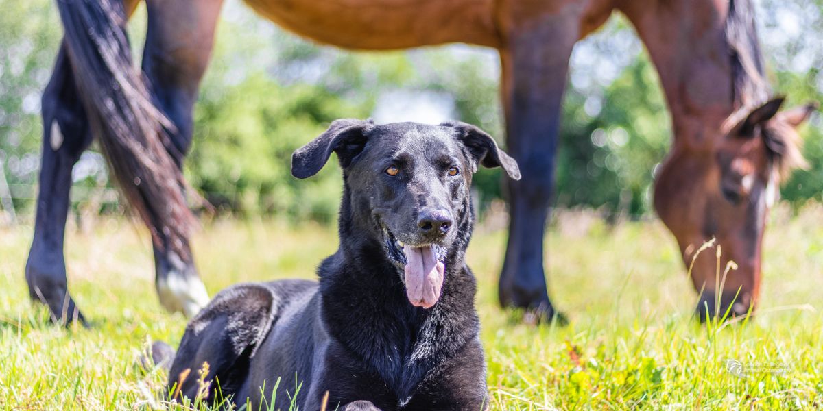
Run a hand down the back of a horse's lower limb, or rest your fingers just above the point of a dog's hock, and the structures you feel there seem almost unremarkable. They are easy to think of as little more than the rope that ties muscle to bone.
That picture undersells them. Tendon function goes well beyond attachment. Some tendons are stiff and position a joint precisely. Others stretch and recoil with every stride, storing energy and giving it back, so that the muscle attached to them has less work to do. How a horse or dog moves depends on these differences, not only on muscle strength or joint range.
Whether you watch animals move for a living or simply want to understand your own horse or dog a little better, knowing how tendons work changes what you notice in a stride.
More Than a Connection: What Tendons Are and How They Transmit Force
Tendons are made mainly of type I collagen, tightly packed and aligned along their length. That collagen is organised in layers of increasing size. Collagen molecules assemble into fibrils, fibrils group into fibres, and fibres are bundled into fascicles. A looser connective tissue called the interfascicular matrix binds the fascicles together. The whole tendon is wrapped in a thin outer layer, the epitenon, and many tendons have a further loose covering outside that called the paratenon. The tendon cells, or tenocytes, sit in rows between the fibres and maintain the matrix around them.
Where a tendon meets its muscle, the junction is called the myotendinous junction. Its attachment to bone is the enthesis, and at many sites the tissue passes through a zone of fibrocartilage before it meets mineralised bone. This gradual change in tissue helps spread stress across the attachment rather than concentrating it along a single line.
How tendons transmit force along the limb
When a muscle contracts, the tendon carries that force to the skeleton, where it either moves a joint or holds it steady. Because the collagen fibres run almost parallel to the tendon's long axis, the tissue is very strong in tension while still flexible enough to curve around joints and bony prominences. Where tendons change direction over a joint, many run through fluid-filled synovial sheaths that let them glide with little friction.
Under a microscope, the collagen fibres of a relaxed tendon show a slight wave known as crimp. The first part of any stretch straightens this crimp out. Once the fibres are straight they bear the load directly, and the tendon becomes much stiffer.
Built for Movement: Positional Tendons and Elastic Energy Storage
Not every tendon is asked to do the same job, and researchers commonly separate them into two broad groups. Positional tendons need to be stiff, because their role is to move and place part of the limb accurately. Energy-storing tendons are more compliant, stretching substantially under load and giving much of that energy back as they recoil (Thorpe et al., 2012).
Stiff tendons for precise placement
The equine common digital extensor tendon is a good example of a positional tendon. It works at low strain, extending the digit and placing the hoof during the swing phase of the stride, and it is not built to bear body weight. What matters in a tendon like this is that muscle shortening translates directly into joint movement, with very little lost to stretch.
Compliant tendons as biological springs
Energy-storing tendons behave quite differently, and biomechanics research describes three ways in which vertebrates exploit them (Alexander, 2002). In running, tendons stretch as the limb takes weight and recoil as it pushes off, storing and returning elastic strain energy and reducing the metabolic cost of each stride. In jumping, a stretched tendon can recoil far faster than muscle is able to shorten, which lets an animal jump further than muscle alone would allow. The third role is less obvious. A springy tendon makes it easier for a muscle to grade the force it produces, but harder to control the exact position of a joint. Elastic storage is not confined to the limbs either, since an aponeurosis (a broad, flat sheet of tendon-like tissue) in the back saves energy at the gallop.
A study of equine tendons found that much of the difference between these two types lies not in the fascicles themselves but in the material between them (Thorpe et al., 2012). In the energy-storing superficial digital flexor tendon, the interface between fascicles was less stiff at physiological loads than in the positional extensor tendon, which allowed the fascicles to slide more freely against one another. That sliding may explain why the whole tendon can stretch further before failing than its individual fascicles can.
Tendons in Action: Equine and Canine Biomechanics
The equine superficial digital flexor tendon
In the horse, the superficial digital flexor tendon (SDFT) is the best-known example of a tendon acting as a spring. There is no muscle in the horse’s lower leg; the superficial digital flexor muscle sits above the knee at the back of the forearm, and its fibres are very short. Below the knee, the tendon itself does nearly all of the stretching and recoiling as the fetlock sinks under load and then rebounds. On the front of the cannon bone, just under the skin, runs the common digital extensor tendon described earlier, so the lower leg holds two tendons with almost opposite jobs on either side of one bone.
The canine common calcaneal tendon
Dogs offer a less familiar but equally instructive example. The common calcaneal tendon, the canine counterpart of the human Achilles, is not a single tendon at all. It brings together the tendon of the gastrocnemius, the superficial digital flexor tendon, which caps the point of the hock before continuing down to the toes, and a contribution from the biceps femoris, semitendinosus and gracilis. Since those last three muscles arise from the pelvis, this one structure links the hip, stifle and hock into a functional chain.
Research on racing greyhounds shows how specialised parts of that chain can become. Williams et al. (2008) found that the gastrocnemius and superficial digital flexor muscles combined short fascicles with long tendons, an arrangement suited to elastic energy storage, while the large hip extensor muscles appeared built for power. Much of the drive comes from the muscles around the hip, while the tendons lower down store energy as the limb takes weight and return it at push-off. A greyhound is an extreme sprinter, though, so these findings show what the canine hindlimb can be adapted for rather than how every dog's limb works.
Why Tendon Anatomy Matters in Movement Assessment
Knowing which tendons store energy and which simply position a joint changes what you notice when an animal moves. In the horse, the fetlock sinks as the limb takes weight and rebounds as it leaves the ground, with the flexor tendons and suspensory ligament working together. In the dog, the hock flexes slightly as weight comes onto the limb and then extends into push-off, loading and unloading the common calcaneal tendon and the muscles attached to it.
The multi-joint nature of these structures matters too. Because the canine common calcaneal tendon draws on muscles that start at the pelvis, a change in how a dog uses its hip may show up at the hock, and the reverse is also possible. Looking at one joint in isolation risks missing where a pattern actually begins.
Tendons in a hands-on assessment
During palpation, tendons are assessed for contour, texture, temperature and sensitivity, and always compared with the same structure on the opposite limb. An osteopathic assessment, including the Osteopathic Articular Balancing (OAB) approach taught at LCAO, looks at how each joint moves in relation to the tissues around it, including the tendons that cross it. Much of this observation starts at walk, and a gradual, walk-first warm-up gives a natural chance to watch, as discussed in our post on warming up a horse before riding.
The Bigger Picture: Clinical Limits and Veterinary Collaboration
Understanding how tendons work is not the same as being able to judge their condition. What happens inside a tendon cannot be reliably seen or felt from the outside. Confirming the state of the tissue needs veterinary examination and imaging, most often ultrasound, and the interpretation of those findings belongs with the vet.
That makes the osteopath's role clear. Heat, swelling, sensitivity or a sudden change in gait are reasons to stop and refer, not to treat. Where a horse or dog is already under veterinary care, osteopathy works within the vet's plan, paying attention to how the rest of the body is coping and reporting back. Our guide to animal osteopath scope of practice sets out how that relationship works, including the requirement in some countries, such as the UK, for the animal's own vet to give permission before treatment.
Tendons rarely get the attention that muscles and joints do, yet they shape how force reaches the skeleton, how precisely a foot is placed and how much energy each stride gives back. Watching a fetlock sink and rebound, or a hock flex and extend, means more once you know what is doing the work.
References
Alexander, R. M. (2002). Tendon elasticity and muscle function. Comparative Biochemistry and Physiology Part A: Molecular & Integrative Physiology, 133(4), 1001–1011.
Thorpe, C. T., Udeze, C. P., Birch, H. L., Clegg, P. D., & Screen, H. R. C. (2012). Specialization of tendon mechanical properties results from interfascicular differences. Journal of the Royal Society Interface, 9(76), 3108–3117.
Williams, S. B., Wilson, A. M., Rhodes, L., Andrews, J., & Payne, R. C. (2008). Functional anatomy and muscle moment arms of the pelvic limb of an elite sprinting athlete: the racing greyhound (Canis familiaris). Journal of Anatomy, 213(4), 361–372.


